Close menu
- Home
- News
- Research
- Opinion
- Features
- Culture
- Careers
- Podcasts
- Webinars
-
Collections
- Back to parent navigation item
- Collections
- The future of energy storage
- AI and automation in chemistry
- Sustainable labs
- Research culture
- Coronavirus
- Nobel prize
-
Themed supplements
- Back to parent navigation item
- Themed supplements
- Problem solvers
- Inspiring science
- Up to the challenge
- Eureka moments
- Chemistry 4.0
- Forefront of pharma
- Collaborative chemistry
- Everyday chemistry
- Future of pharma
- Voices in chemistry
- Chemistry detectives
- Innovators
- Green and sustainable chemistry
- Health technology
- View all
- Partner collections
- Register


A slice of ion beam–scanning microscopy
By Emma Davies2017-06-14T13:47:00
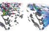
Source: © Bert Weckhuysen / Utrecht University
From brain cells to batteries, is there anything focused ion beam–scanning electron microscopy can’t study?
Focused ion beam–scanning electron microscopy (FIB–SEM) was originally targeted at the semiconductor industry but is now used in an astonishing array of applications. Each technique has been used separately for decades but researchers are increasingly drawn to the combined technique, which brings both high-resolution imaging and micromachining.
- This website collects cookies to deliver a better user experience. See how this site uses cookies.
- This website collects cookies to deliver a better user experience. Do not sell my personal data.
- Este site coleta cookies para oferecer uma melhor experiência ao usuário. Veja como este site usa cookies.
Site powered by Webvision Cloud